
German researchers develop new software for virtual exploratory tours in a network of tiny blood vessels
Researchers at the Universities of Bayreuth and Marburg in Germany have succeeded in creating high-definition images of human blood vessels magnified thousands of times. Using virtual reality glasses from the world of computer games, it is now possible to take virtual exploratory tours through a complex mesh of tiny blood vessels. In this way, scientists were able to make new discoveries about the spleen, which they have now published in the journal PLOS ONE.
In 2017, research teams led by Prof. Dr. Michael Guthe and Dr. Oleg Lobachev in Bayreuth (Computer Science) and Prof. Dr. Birte Steiniger in Marburg (Anatomy) created a method that enables them to derive high-definition 3D models of the tiny blood vessels in bone marrow and spleen. On this basis, they have now developed new software that enables the images of tissue sections to be put together to form 3D models at about thousandfold magnification. Virtual reality glasses, like those used in certain computer games, take the observer on a trip through networks of blood vessels, many of which are only a few thousandths of a millimetre wide. This allows blood vessels to be examined in much greater detail than was previously possible. At the same time, the images of the tissue sections can be superimposed on the resulting magnified models. This enables direct comparisons that facilitate the ongoing quality control of the models.
The computer scientists in Bayreuth emphasize that this virtual reality technology is particularly revealing in the area of basic medical research. Routinely employing the technology in diagnostics, however, requires faster computers capable of processing huge amounts of data in a short time.
New findings concerning blood vessels in the spleen
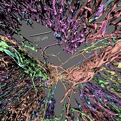
The research groups led by Prof. Dr. Michael Guthe and Dr. Oleg Lobachev in Bayreuth (Computer Science) and Prof. Dr. Birte Steiniger in Marburg (Anatomy) have applied the new visualization process to clarify the structure and path of tiny vessels known as capillaries in the human spleen. What is fundamental in this connection is the division of the spleen into two areas, the white and red pulp, which perform different functions. The white pulp contains mainly two different types of white blood cells: B and T lymphocytes. It has very few capillaries. At the surface of the white pulp – particularly at the surface of B lymphocyte clusters known as follicles – a tight network of capillaries can be found. These are mainly supplied by a more coarsely structured capillary network of the surrounding red pulp.
At the surface of the follicles and in the red pulp, many capillaries have open ends. “Our 3D models thus enable one to clearly see how the spleen is involved in blood circulation in the human body. The blood appears to flow from the open capillary ends in the splenic red pulp without being contained in vessels, before it re-enters the venous circulation. Here it comes into direct contact with macrophages, which free the blood of harmful foreign materials and aged red blood cells,” Dr. Oleg Lobachev explained.
 How to resolve AdBlock issue?