BIG DATA
FAU prof Kainz wins 2 million euros for ML in medical imaging diagnostics

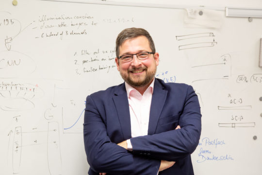
It is a feather in the cap for outstanding research at the Department of Artificial Intelligence in Biomedical Engineering at Friedrich-Alexander-Universität Erlangen-Nürnberg (FAU) in Bavaria, Germany: The European Research Council (ERC) has awarded a Consolidator Grant to Prof. Dr. Bernhard Kainz. The professor of Image Data Exploration and Analysis has received the most prestigious research funding award available throughout the whole of Europe for a project focusing on automated medical image analysis. The two million euros in funding over five years is to be used to train computer tools based on artificial intelligence to reliably recognize healthy human tissue based on image material. The aim is to use these tools for medical diagnosis to support and ease the workload of experts working in the field, for example during health screening programs.
Improving medical care
Imaging is playing an increasingly significant role in medicine, but analyzing images is both time-consuming and costly. It not only ties up existing resources and leads to an increase in costs, but it also causes long waiting times for patients. With their machine learning project, Professor Kainz and his team hope to train computer programs to recognize healthy tissue structures. Artificial intelligence would then be able to pre-sort the images obtained during the diagnosis process into “probably healthy” or “possibly sick”. The final decision is taken by medical experts. With the support of the machines, however, medical staff would gain valuable time that they could then use to investigate any images deviating from the norm more thoroughly. As a knock-on effect, more patients could be treated, and patients would not have to wait so long to find out whether the images indicated that there was a problem with their health.
According to Bernhard Kainz, he is driven by the “conviction that everyone deserves the same quality of medical care, no matter where they live or how much they earn. That is why our working group is working on developing methods that make high-quality medical imaging analyses widely available and scalable.”
Recognize healthy tissue
Why should AI recognize healthy tissue? Why is it not being trained to diagnose diseases? Prof. Kainz has a clear answer: “Training machine learning tools using hundreds and hundreds of examples of every possible disease would be extremely costly in terms of time and manpower. Medical experts who are already overstretched would have to provide and comment on vast amounts of images of pathological structures.” In his opinion, it makes much more sense to “feed” the AI with images of healthy tissue structures. That is time-consuming enough, as healthy tissue differs depending on age and other characteristics such as gender.
The team is therefore working on providing self-learning machine diagnostic tools that recognize what healthy anatomy should look like at a certain point in time. The computer tools are to be trained to recognize normal physiological traits and any unusual changes over a certain period in individual patients. In addition, they should match patient information provided by physicians (for example laboratory results) to the available images. Bernhard Kainz’s goal is that the tools will recognize any deviations that require a more detailed medical investigation, at the same time as avoiding any unnecessary examinations, for the benefit of the patients.
